Single Molecule Live Cell Imaging

Seeing is believing...
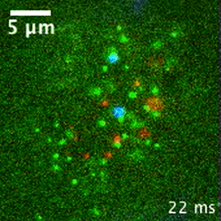
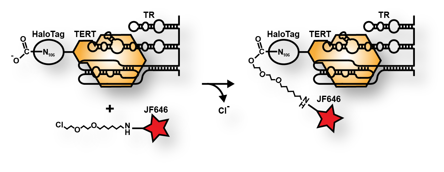
Single molecule imaging is a powerful technique. It allows the observer to measure the behavior of individual molecules rather than the average of the whole population. For instance, it revealed to us that telomerase in the movie above in green, behaves differently at telomeres (red) and Cajal bodies (blue) than at other nuclear locations. To visualize single proteins in living human cells we take advantage of the HaloTag, a small protein, that can be covalently modified with a cell permeable small molecule fluorophore. We expose cells expressing a HaloTagged protein from its endogenous locus to a small dose of fluorophore for a short time to label only a subset of the molecules in the cell. Using this approach we achieve a particle density that allows the detection of single molecules, without much overlap between the fluorescence signals. We visualize the fluorescent molecules using a TIRF microscope equipped with powerful lasers and EMCCD cameras and we can image multiple channels with high temporal resolution (50 frames per second). To aid the detection of single molecules we generate a thin light sheet through the sample, to reduce out of focus fluorescence, a technique called Highly Inclined Laminated Optical Sheet (HILO) microscopy. If you are interested in learning more about this technology, or are potentially interested in setting up a collaboration, please contact us.